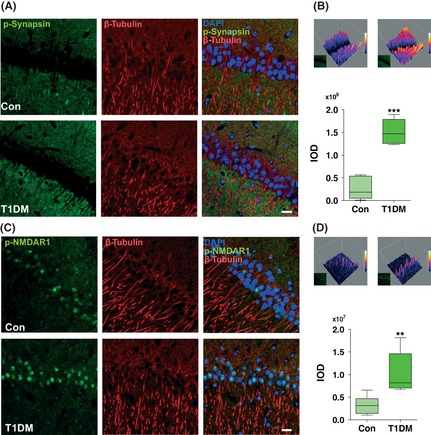
Figure 5.

The STZ treatment induced aberrant immunostaining of phospho‐synapsin I (Ser 603) and phospho‐NMDAR1 (Ser 896) in type 1 diabetic rats. Immunohistochemical localization of phospho‐synapsin I (Ser 603) (A, B) and phospho‐NMDAR1 (Ser 896) (C, D) was examined in the hippocampus CA1 region. Markedly increased phospho‐synapsin I (Ser 603) (A) and phospho‐NMDAR1 (Ser 896) (C) stainings were observed by confocal microscopy in the hippocampus of type 1 diabetic rats. DAPI counterstaining indicates nuclear localization (blue). Scale bar = 50 μm. The 3D filled surface plots (Upper) indicate the grayscale intensities of phospho‐synapsin I (Ser 603) (B) and phospho‐NMDAR1 (Ser 896) (D). The integrated optical density (IOD) determined by Graphpad Prism v5.0 according to the staining intensity of proteins (Lower). **P < 0.001; ***P < 0.001 versus control (n = 6 slices from three animals for each group). T1DM, type 1 diabetes mellitus.
