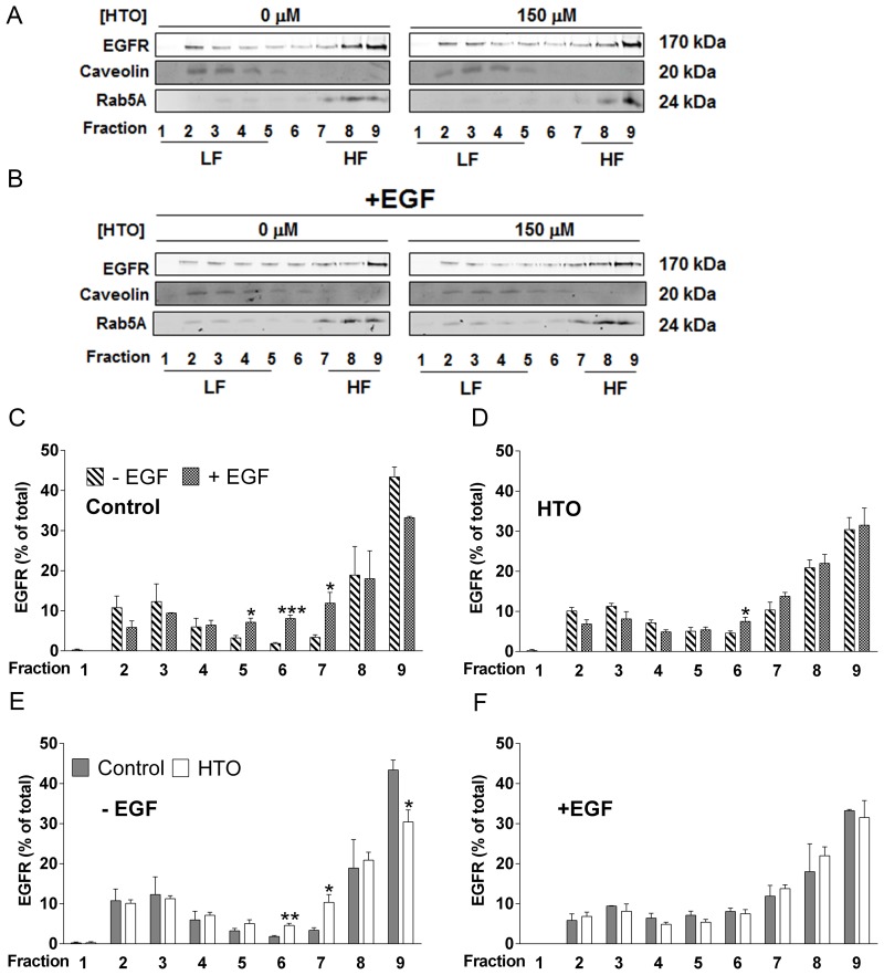
Figure 5. Effect of HTO on the membrane microdomain distribution of EGFR.
(A–F) MDA-MB-231 cells were serum starved and cultured for 24 h in the presence or absence of HTO (150 μM), before they were exposed to EGF (100 ng/ml) or the vehicle alone for 30 min. The cells were collected, solubilized in 1% Brij 98 polyoxyethylene fatty ether detergent and subjected to sucrose gradient separation before analyzing the EGFR distribution in detergent-resistant heavy (HF) and light (LF) membrane fractions in western blots (identified by Rab5 and Caveolin, respectively). Representative western blots are shown and the bars correspond to the mean ± SEM values of 2 independent experiments: *p < 0.05, **p < 0.01, ***p < 0.001 vs control.

