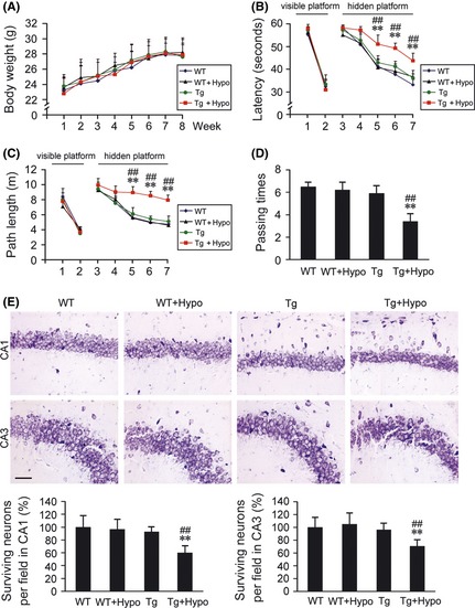
Figure 1.

Effects of hypoxia treatment on spatial learning and memory, and the hippocampal neuronal apoptosis. (A) During 2 months hypoxia intervention, the body weight evaluation of hypoxia‐treated transgenic APP/PS1 mice (Tg + Hypo) and age‐matched wild‐type C57BL/6 mice (WT + Hypo) or the groups under control (Tg or WT), respectively. (B–D) Morris water maze tests showed the spatial learning and memory capabilities of these four groups. Mice exhibited the similar escape latency and path length in the visible platform training from day 1st to 2nd, whereas, from day 5th to 7th, mice in Tg + Hypo group performed the longest latency and escape length in hidden platform tests. In the probe trial on the 8th day, the mice in Tg + Hypo group exhibited the less passing times into the northwest quadrant, where the platform was previously located; All values are mean ± SEM (n = 6); **P < 0.01 versus WT group, ## P < 0.01 versus Tg group (repeated measures ANOVA). (E) Nissl staining showed the surviving neurons (round and palely stained nuclei) and the apoptosis neurons (shrunken neurons with pyknotic nuclei) in the CA1 and CA3 of the hippocampus. Scale bar = 20 μm. All values are mean ± SEM (n = 6); **P < 0.01 versus WT group, ## P < 0.01 versus Tg group (two‐way ANOVA was adopted for factorial designs).
