Figure 2.

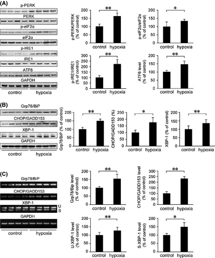
Hypoxia induces the activation of UPR and increases the expression of ER‐resident chaperones in the APP/PS1 mouse brain. Six‐month‐old female APP/PS1 transgenic mice were exposed to hypoxic conditions once daily for 2 months. UPR signaling and ER stress–response proteins were analyzed by Western blot and RT‐PCR. GAPDH was used as an internal control. (A) The increase in p‐PERK/PERK, p‐eIF2α/eIF2α, and p‐IRE1/IRE1 in the APP/PS1 mouse brain exposed to hypoxic conditions indicated the activation of PERK, eIF2α, and IRE1, respectively. In addition, Western blot analysis of ATF6 (active form of ATF6, 50 kDa) showed a significant increase in the hypoxia group. (B) The protein level of ER‐resident chaperones Grp78/BiP was markedly increased, and CHOP/GADD153 and XBP‐1 expression levels were significantly elevated by hypoxia treatment, compared with the control. The changes in mRNA levels of Grp78/BiP and CHOP/GADD153 detected by RT‐PCR were in agreement with the protein expression analysis. RT‐PCR showed the levels of unspliced (U) and spliced (S) XBP‐1. (C) Analysis of XBP‐1 mRNA splicing indicated that hypoxia treatment significantly increased the presence of spliced XBP‐1 mRNA in the APP/PS1 mouse brain. All values are mean ± SEM (n = 6); *P < 0.05, **P < 0.01 versus control group (Student's t‐test).
