
Figure 6.
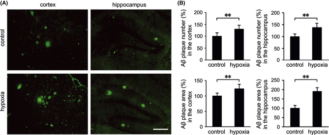
The effects of hypoxia on Aβ plaque deposition in the APP/PS1 mouse brain. (A) Brain sections of APP/PS1 mice from the control and hypoxia groups were labeled with anti‐Aβ to detect Aβ plaques by immunofluoresence. (B) Quantification shows that more Aβ plaques were stained in the cortex and hippocampus of the hypoxic mouse brain. Scale Bar = 100 μm. Values are mean ± SEM (n = 6); **P < 0.01 versus control group (Student's t‐test).