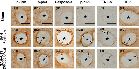
Figure 4.

Immunohistochemical staining of the microvasculature in hippocampus. The levels of phosphorylated‐JNK, phosphorylated‐p53, caspase‐3, phosphorylated‐NF‐κB (p65), tumor necrosis factor‐α (TNF‐α), interleukin‐6 (IL‐6) in endothelial cells of microvasculature in hippocampus were markedly elevated, in addition, the numbers of endothelial cells with positive staining were also increased (A1–F1, A2–F2).These pathologies were significantly attenuated by 50,000 U/kg urinary trypsin inhibitor (UTI) treatment (A3–F3). Scale bars = 10 μm, “stars” indicated microvessels; “arrows” showed the endothelial cells, n = 6 each group.
