
Figure 5.
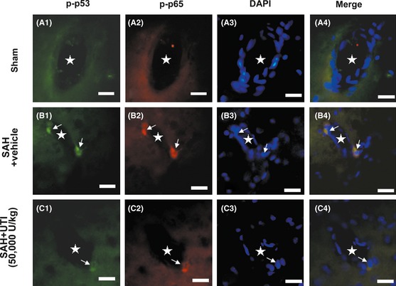
The immunofluorenscence staining of the microvasculature in hippocampus. In sham group, the phosphorylated‐p53 and NF‐κB (p65) were bare in the endothelial cells (A1–A4). After subarachnoid hemorrhage (SAH), p53 and NF‐κB (p65) in the endothelial cells were simultaneously activated and distributed in the nucleus (B1–B4). Urinary trypsin inhibitor (UTI) treatment could significantly suppress the levels of phosphorylated‐p53 and NF‐κB (p65) in the endothelial cells (C1–C4). Scale bars = 20 μm, “stars” indicated microvessels; “arrows” showed the endothelial cells, n = 6 each group.