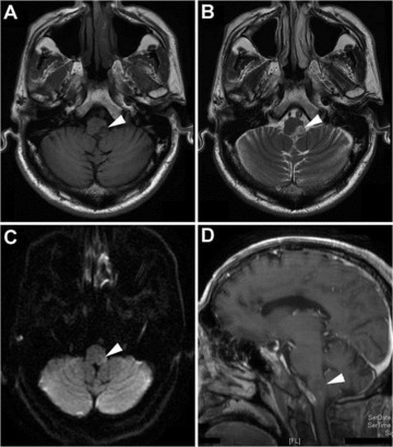
Figure 1.

MR imaging obtained 16 days after symptom onset. MR image through the medulla oblongata at presentation shows a patchy hypointense (A), hyperintense (B and C), and nonenhancing (D) lesion over the left posterolateral aspect of the medulla oblongata (arrow heads) on T1‐weighted (A), T2‐weighted (B), diffusion‐weighted (C), and enhancing (D) imaging, respectively.
