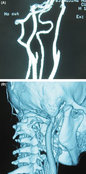
Figure 2.

The cervical 3D‐computed tomography angiography (CTA) and 3D‐CT of the patient 3 months after the symptoms onset. (A) The cervical 3D‐CTA showed a smooth recanalized and free‐flowing right VA without thrombosis. (B) Cervical 3D‐CT and 3D‐CTA shows the transverse foramen of C1 moved forward as a result of atlas dislocation. This led to the foramina of C1 and C2 retracting the VAs and made them compressed around the C2 foramen (↑).
