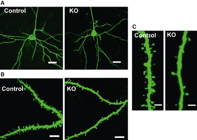
Figure 2.

Morphological changes in the prefrontal cortex of ventral forebrain‐specific heparin‐binding epidermal growth factor (HB‐EGF) knockout (KO) mice. (A) Representative photomicrographs showing morphology of pyramidal neurons in cortical layer III of the prefrontal cortex from wild‐type control (left) and KO (right) mice. Scale bar = 20 μm. (B) Representative photomicrographs of apical dendritic segments from wild‐type control (left) and KO (right) mice. Scale bar = 8 μm. (C) High‐magnification images of apical dendritic segments from wild‐type control (left) and KO (right) mice. Scale bar = 2 μm. These figures were reproduced from Oyagi et al. 51.
