
Figure 2.
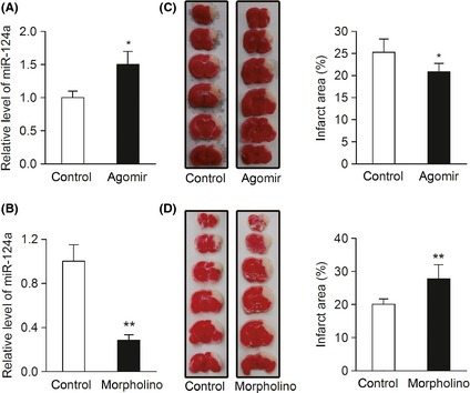
MiR‐124 protects against acute ischemic cerebral injury in MCAO mice. (A, B) Real‐time PCR detection of miR‐124 in the brain tissues of C57BL/6 mice 3 days after tail‐vein injection with miR‐124 agomir, vivo‐morpholino, and their matched controls. (n = 3, *P < 0.05, **P < 0.01). (C) C57BL/6 mice were injected via tail‐vein with 100 nmol/kg of miR‐124a agomir or negative control for 3 days, and then underwent MCAO by electrocoagulation. 24 h after MCAO operation, infarct area was evaluated by TTC staining. (n = 7, *P < 0.05). Left, representative images of TTC‐stained brain sections; right, quantitative analysis of infarct area. (D) C57BL/6 mice were injected via tail‐vein with 2 mg/kg of antisense miR‐124 vivo‐morpholino or control vivo‐morpholino for 3 days, and then treated as described in (C). (n = 8, **P < 0.01). Data are represented as mean ± SD.