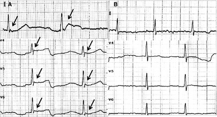
Figure 1.

A 12‐lead electrocardiogram (ECG) was taken immediately after admission (when aural canal temperature was 30.9°C) and showed the obvious Osborn wave on lead II and lead V4–V6 (black arrow in A). The repeated ECG taken after the patient's body temperature returned to normal (aural canal temperature was 37.0°C) revealed no Osborn waves (B).
