
Figure 5.
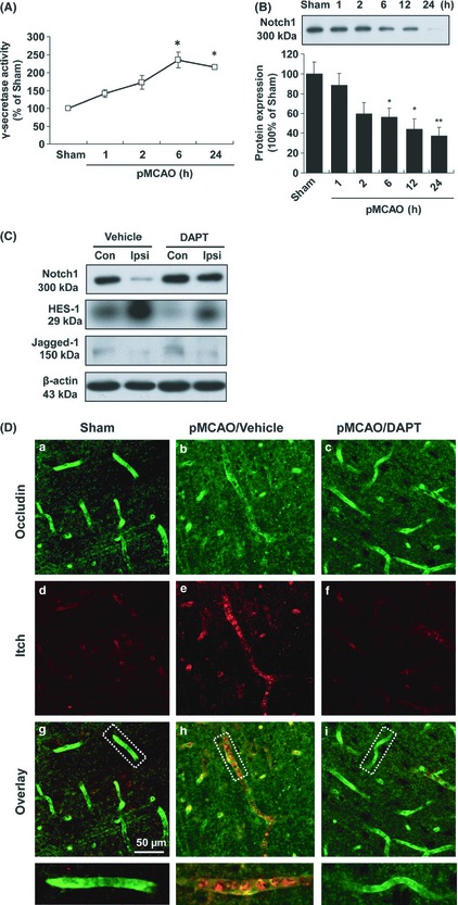
The neurovascular protective effect of DAPT is associated with its inhibitory effect on γ‐secretase and Itch activation in pMCAO rats. (A) The changes in γ‐secretase activity in the rat brain following pMCAO. Data are presented as mean ± SEM (n = 3); *P < 0.05 versus sham‐operated rats. (B) Representative immunoblots probed with an anti‐Notch1 antibody. A 300‐kDa Notch1 band was observed in the brain extracts from sham and pMCAO rats. Data are expressed as the percentage of the value observed in sham‐operated animals (mean ± SEM, n = 4). *P < 0.05 versus sham‐operated rats. (C) Representative immunoblots from brain lysates of vehicle‐ and DAPT‐treated pMCAO rats when probed with anti‐Notch1, ‐HES‐1 and ‐Jagged‐1 antibodies. Con, contralateral hemisphere; Ipsi, ipsilateral hemisphere. (D) Fluorescent staining of occludin and Itch in the cerebral microvessels of pMCAO rats. Anti‐occludin (a, b, and c) and ‐Itch (d, e, and f) staining was performed 24 h after pMCAO with or without DAPT treatment. Scale bar = 50 μm.