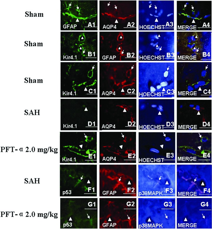
Figure 5.

Treble immunofluorescence staining of the microvessels in rat hippocampus. In the sham‐operated animals, the treble fluorescence labeling of GFAP, AQP4 and Hoechst 33258 (for nuclear) (Figure A1–A4) and Kir4.1, GFAP and Hoechst 33258 (Figure B1–B4) and Kir4.1, AQP4 and Hoechst 33258 (Figure C1–C4) showed that both AQP4 and Kir4.1 were mainly distributed on the end feet of astrocytes and along the walls of the microvessels. Following SAH, the expression level of Kir4.1 was markedly declined and whereas AQP4 was elevated, which indicated that there was uncoupling expression between Kir4.1 and AQP4 in the astrocytes end feet (Figure D1–D4). In addition, activated p53 and phosphorylated‐p38MAPK were colocalized in the astrocytes whose end feet terminated on the wall of the microvessls (Figure F1–F4). Both tendencies in figures D, F after SAH could be reversed by PFT‐α 2.0 mg/kg treatment (Figures E and G). Scale bars = 40 μm in Figures C and E, scale bars = 20 μm in other figures. “Triangles” indicated microvessels; “arrows” showed the astrocytes.
