
Figure 1.
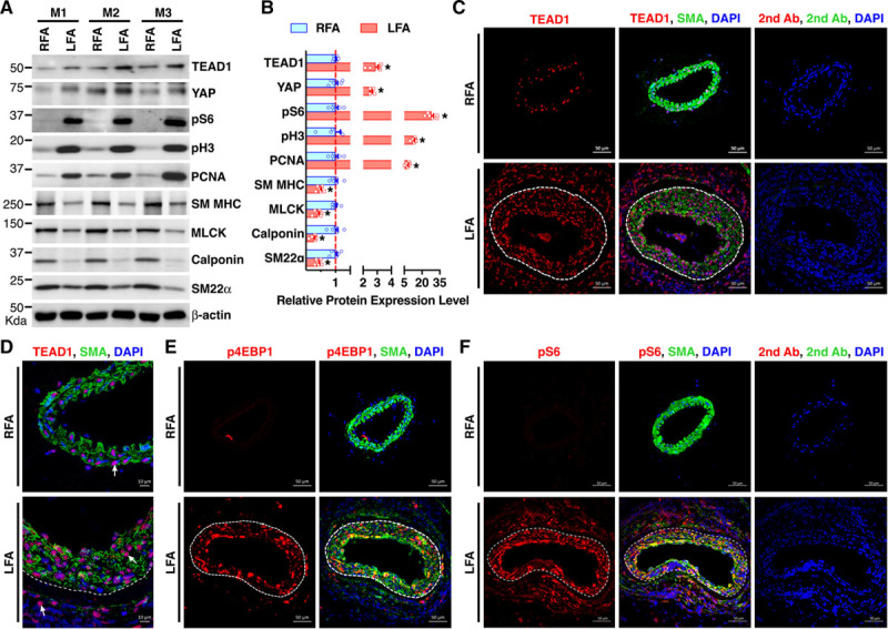
TEAD (TEA domain transcription factor) 1 expression is induced after arterial injury and positively correlates with mTORC1 (mechanistic target of rapamycin complex 1) activation in vascular smooth muscle cells in vivo. A, Control right femoral arteries (RFAs) or wire-injured left femoral arteries (LFAs) were harvested from adult male C57BL/6 mice (M) 7 d post-injury and analyzed by Western blot. B, Densitometric analysis of protein expression in A normalized to the loading control (β-actin) and expressed relative to signals from the uninjured control RFA (set to 1, red line). C and D, Control RFAs or wire-injured LFAs were harvested from adult male C57BL/6 mice 14 d post-injury and analyzed by immunofluorescence (IF) staining for TEAD1 (red) or SMA (smooth muscle α-actin; green). Nuclei were counterstained with DAPI (4’,6-diamidino-2-phenylindole; blue). Representative TEAD1 staining is shown by arrows (D). E and F, IF was performed similar to C except for using antibodies for activated mTOCR1 markers (E) p4EBP1 (phospho-eukaryotic initiation factor-4E–binding protein-1) or (F) pS6 (phospho-ribosomal protein S6), respectively. White dashed line denotes internal elastic lamina. The sections stained with fluorophore-labeled secondary antibodies (2nd Ab) and DAPI served as negative control. MLCK indicates myosin light-chain kinase; SM MHC, smooth muscle myosin heavy chain; and SM22α, smooth muscle protein 22-alpha. *P<0.05.