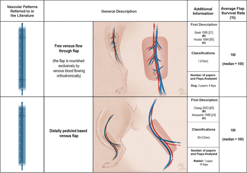
Fig. 4.

Schematic representation of the free venous flow-through flap and the distally pedicled venous flap performed in experimental models according to the literature. These flaps receive venous blood through one or more of their veins. Drainage of venous flaps occurs through one or more veins to neighboring veins. Red areas represent arterial blood flow. Blue and purple regions denote venous and mixed arterial and venous blood, respectively. The arrows specify the direction of blood flow. The curved lines inside the vessels illustrate venous valves. First description: in cases where the first description of the type of unconventional pattern was not performed in the experimental setting (E), the description in the clinical setting (C) is also indicated. Classifications: The classifications used were those proposed by Woo et al. (Woo SH, Kim KC, Lee GJ, et al. A retrospective analysis of 154 arterialized venous flaps for hand reconstruction: An 11-year experience. Plast Reconstr Surg. 2007;119:1823–1838) and by Chen et al. (Chen HC, Tang YB, Noordhoff MS. Four types of venous flaps for wound coverage: A clinical appraisal. J Trauma 1991;31:1286–1293). The drawings are not to scale.
