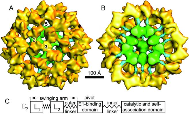
Figure 2.
3D reconstruction of bovine PDC (A and B) and diagrammatic representation of the structural domains of E2 subunit (C). Shaded-surface representation of 3-fold axes of symmetry of the 3D reconstruction of the bovine kidney PDC (A) and with the closest half removed to reveal the linker (blue) that binds E1 (yellow) to the E2 core (green; B). The inner linker is ≈50 Å in length. (C) The C-terminal self-association domain is responsible for the assembly of the dodecahedral scaffold to which E1 and BP⋅E3 bind. The N-terminal half of the E2 comprises the L1 and L2 lipoyl domains, and the E1-binding domain, and their associated linkers. The inner linker is revealed in the 3D structure of the PDC.

