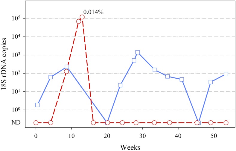
Figure 2.
Time course of Plasmodium malariae infections. Shown are the copy numbers of Plasmodium 18S rDNA detected in each QMAL reaction (equivalent to 8 μL of blood). Blue solid line with squares, 1st participant; red dashed line with circles, 2nd participant. 18S rDNA copy below 100 considered as QMAL-negative samples in which parasite was ND; 0.014% indicates the parasitemia of participant 2 at the clinical passive case detection visit on August 20, 2013, as determined by light microscopy. ND = not detected; QMAL = genus-specific quantitative PCR assay. This figure appears in color at www.ajtmh.org.

