Figure 1.
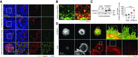
Thrombospondin type 1 domain–containing 7A (THSD7A) expression is a characteristic of mature podocytes. (A) Representative confocal micrographs of THSD7A (green) expression in the postnatal day 5 kidney of a Balb/C mouse using the goat anti-THSD7A antibody. Nephrin (red) was used to visualize premature to mature podocytes, Lotus tetragonolobus lectin (LTA; light blue) marks the glycocalyx of proximal tubuli brush border, DNA is in blue. (B) Decapsulated murine glomeruli were isolated and plated onto collagen type IV–coated plates for 4 days. Primary glomerular epithelial cells (GECs) that have grown out of the tuft were stained for THSD7A (green) and nephrin (red). The asterisk labels the glomerular tuft from which the GECs have grown out. Note the decrease in THSD7A expression in cells that have migrated farther away from the glomerular tuft (arrows), whereas nephrin immunoreactivity is maintained. (C) Western blot analyses of murine podocytes cultured under permissive (P; 32°C) and nonpermissive (NP; 38°C) conditions against murine THSD7A. β-actin of the same samples run under reducing WB conditions was used to control for equal loading. The graph exhibits densitometric analyses of three independent experiments; values are expressed as median, and they are calculated as percentage change to permissive mock levels (P-mock). ***P<0.001 (one-way ANOVA, Dunnett multiple comparison test). (D) Murine immortalized podocytes were transduced with murine thrombospondin type 1 domain–containing 7A (mTHSD7A) or control transduced (mock) with empty vector. Representative confocal micrographs of THSD7A (green) expression in relation to filamentous actin (F-actin; red) are shown. Note the membranous expression of THSD7A, which is also in thin actin free processes (arrows).

