Figure 2.
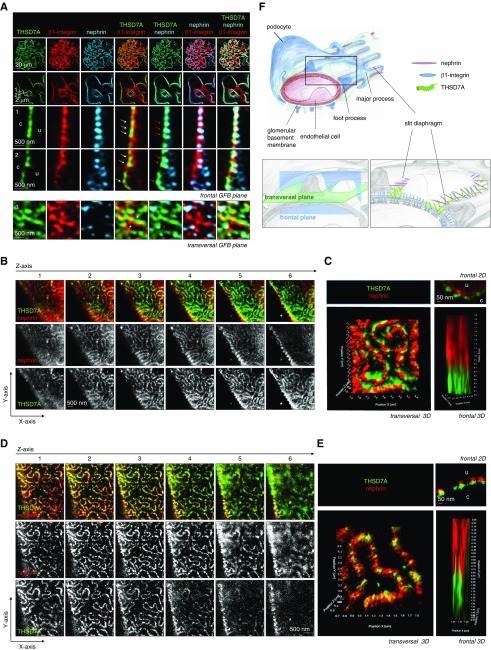
Thrombospondin type 1 domain–containing 7A (THSD7A) localizes to podocyte foot process domains close to the slit diaphragm. (A) Three-micrometer-thick paraffin sections of a naive C57BL/6 mouse were stained for THSD7A (green), β1-integrin (red), and nephrin (light blue). The glomerular filtration barrier (GFB) is visualized in the enlarged boxes in the frontal plane or the transversal plane as indicated. Note the localization of THSD7A side by side in an alternating pattern with β1-integrin (white arrows) at the basal aspect of foot processes (FPs). Also note the localization of THSD7A basally of nephrin in the frontal plane (red arrows) but in the same FP domain when visualized in the transversal plane. c, capillary side of GFB; u, urinary side of GFB. (B) Z stacks of THSD7A (green) and nephrin (red) localization visualized by stimulated emission depletion (STED) microscopy of a naive C57BL/6 mouse paraffin section. (C) A 3D reconstruction of THSD7A (green) and nephrin (red) localization in the same FP domain of the slit diaphragm but with THSD7A located basally from nephrin as visualized by STED microscopy of a naive C57BL/6 mouse paraffin section. (D) Z stacks of THSD7A (green) and nephrin (red) localization visualized by STED microscopy of a human frozen section derived from the healthy part of a tumor nephrectomy. (E) A 3D reconstruction of THSD7A (green) and nephrin (red) localization in the same FP domain of the slit diaphragm but with THSD7A located basally from nephrin as visualized by STED microscopy of a human frozen section derived from the healthy part of a tumor nephrectomy. (F) A 3D scheme of GFB indicating the localization of the frontal versus the transversal optical plane (lower left panel) and the proposed localization of THSD7A in relation to β1-integrin and nephrin.

