Figure 6.
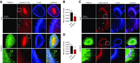
Thrombospondin type 1 domain–containing 7A (THSD7A) expression in podocytes dampens activity of cytoskeletal regulatory proteins in differentiated cultured human podocytes. (A) Representative confocal micrographs of THSD7A (green), the activated (Y118-phosphorylated) form of the focal adhesion protein paxillin (which is involved in integrin-mediated cytoskeletal reorganization), and filamentous actin (F-actin; blue). Note the prominent localization of p-paxillin at focal adhesions (arrows) in the mock-transduced control podocyte, which is nearly absent in the THSD7A-expressing podocyte. (B) Quantification of mean intensity of fluorescence (MIF) of p-paxillin per cell normalized to cell area in n=10 cells of three independent experiments; values are depicted as the mean±SEM. *P<0.05 (unpaired t test). (C) Representative confocal micrographs of THSD7A (green), the activated (Ser19-phosphorylated) form of myosin light chain (MLC; which is required for the formation of crossbridges to actin for contraction), and F-actin (blue). Note the presence of p-MLC at actin fibers in the mock podocyte (arrows) and the prominent cortical p-MLC ring (red arrows) in the THSD7A-expressing podocyte. (D) Quantification of MIF of p-MLC per cell normalized to cell area in n=5–8 cells of three pooled independent experiments; values are depicted as the mean±SEM. hTHSD7A, human thrombospondin type 1 domain–containing 7A. *P<0.05 (unpaired t test).

