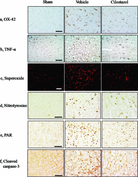
Figure 2.

Representative immunohistochemical staining of the CD11b (OX‐42), TNF‐α, superoxide, nitrotyrosine, poly(ADP‐ribose) polymer (PAR), and cleaved caspase‐3‐positive cells in the penumbral region of the vehicle‐ and cilostazol‐treated group in comparison with sham group. Compared with the vehicle group, the OX‐42, TNF‐α, superoxide, nitrotyrosine, PAR, and cleaved caspase‐3‐positive cells in the penumbral region of the cilostazol‐treated group are less prominent at 24 h reperfusion after 2 h MCAO. Little immunoreactivity was shown in the sham groups. Each representative Figure is derived from 4∼6 rat brains of each group. Scale bar = 50 μm.
