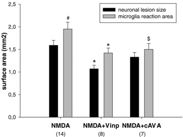
Figure 5.

Extent of neuronal lesion and the area of microglia activation in the entorhinal cortex‐hippocampus complex following NMDA injections at the –7.6 Bregma level. Groups contained 7–14 animals. Vinpocetine (Vinp) treatment reduced the size of both parameters: *P < 0.05 versus NMDA‐vehicle‐treated control group. Treatment with cis‐apovincaminic acid (cAVA) did not diminish significantly the neuronal lesion size, while practically significantly reduced the area covered by activated microglia ($P= 0.05 vs. NMDA‐vehicle group). Area invaded by activated microglia extended the size of neuronal lesion but only in case of NMDA‐vehicle group (# P < 0.05).
