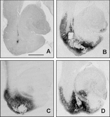
Figure 7.

Areas of microglial activation in the entorhinal cortex‐ventral hippocampus region outlined by DC11b immunostaining. Photomicrographs are from representative animals of sham‐lesioned vehicle‐treated (A), NMDA‐lesioned vehicle‐treated (B), NMDA‐lesioned vinpocetine‐treated (C), and NMDA‐lesioned cAVA‐treated (D) groups. Bar represents 1 mm.
