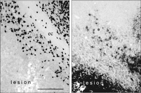
Figure 8.

Photomicrographs with higher magnification allow to identify the stained neurons and microglia cells. The left panel shows the neuron‐specific nuclear protein (NeuN) positive neuronal staining, the right panel the CD11b positive microglia staining. It is noteworthy that the border of neuronal lesion and the microglia activation is sharp and can be clearly outlined. In some cases a scattered appearance of activated microglia could also be observed (asterisks). S, subiculum; ec, external capsule. In the external capsule no neuronal cell bodies are stained. Bar represents 100 μ.
