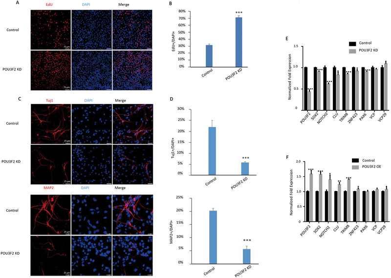
Fig. 5. POU3F2 regulates proliferation and differentiation of NPCs.
(A, B) Immunofluorescence staining for EdU (a marker of proliferating cells) after POU3F2 knockdown in human NPCs (A); quantification of proliferation is shown in panel B. (C, D) Immunofluorescence staining for Tuj1 (a marker of immature neurons) and MAP2 (a marker of mature neurons) after POU3F2 knockdown in NPCs (C); quantification of differentiation of NPCs into neurons is shown in D. (E, F) qPCR data showing POU3F2 putative targets after knocking down (E) or overexpressing POU3F2 (F). Three biological replicates were used, and for each biological replicate we designed three technical replicates. * P < 0.05, ** P < 0.01, ***P < 0.001. Data are represented as mean ± SEM.

