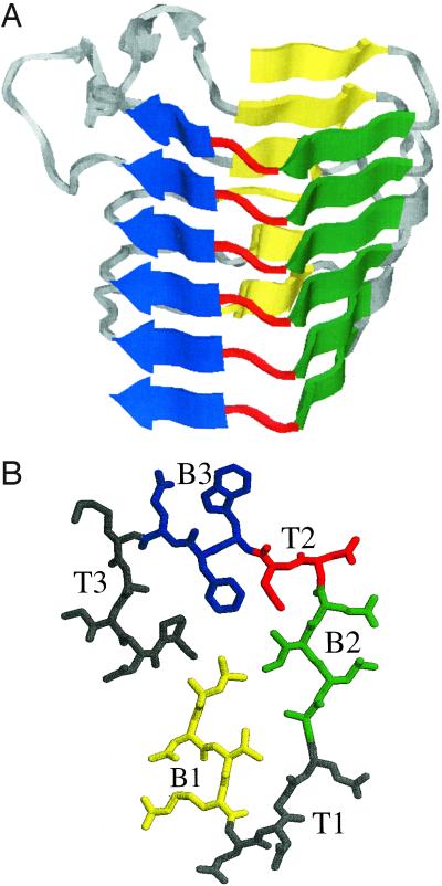
Figure 1.
(A) Side view of x-ray crystal structure (1) of Pectate lyase C from Erwinia chrysanthemi, residue 102–258, generated using the molecular graphics program rasmol (29). (B) Top view of a single rung of a β-helix (residues 242–263 of A), with β-strands B1 in yellow, B2 in green, and B3 in blue, and the intervening turns T1, T2 (in red), and T3. The alternating pattern of the strands before and after T2 and the T2 turn itself are conserved across the superfamily (3, 8).

