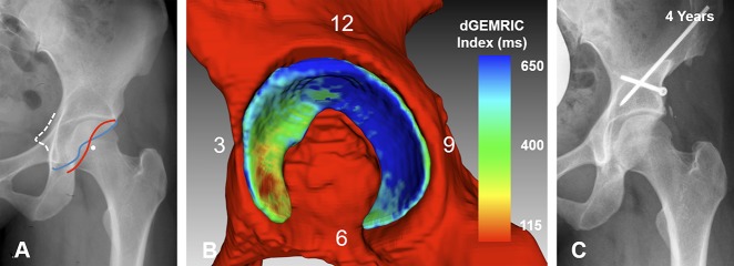
Fig. 7 A-C.
(A) An AP pelvis image shows a 27-year-old woman with severe acetabular retroversion (positive cross-over sign, posterior wall sign, and ischial spine sign). (B) Fully-automated 3-D cartilage model is shown. The dGEMRIC indices are color-coded. Biochemical cartilage damage as shown with lower (green) dGEMRIC indices is present anteriorly compared to the high dGEMRIC indices posteriorly (blue). The overall (1250 mm2) and the anterosuperior cartilage surface area were smaller (457 mm2) compared with the respective means of the entire study group (overall: 1617 ± 426 mm2; anterosuperior: 610 ± 204 mm2; Table 7). The patient underwent anteverting periacetabular osteotomy for correction of malrotation of the hemipelvis. (C) AP pelvis view at the last clinical followup 4 years after surgery shows no progression of osteoarthritis and the patient was asymptomatic.

