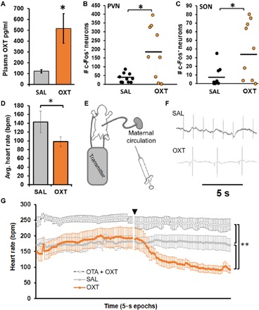
Fig. 1. The fetus is acutely responsive to maternal OXT.

(A) Experiment 1a: Plasma OXT was higher in fetal vole pups after maternal treatment with OXT (0.03 mg/kg, n = 6) compared to saline vehicle (SAL, n = 7). Results from two OXT litters were discarded because of excessively high values that fell beyond the linear portion of the assay’s sensitivity curve. (B and C) Experiment 1b: In a separate set of animals, the number of c-fos + nuclei was higher in fetal vole pups after maternal treatment with OXT (0.03 mg/kg, n = 9) compared to SAL (n = 10) in both the paraventricular nucleus (PVN) (B) and supraoptic nucleus (SON) (C). (D) Experiment 2a: Heart rate in postnatal day 1 (PND 1) vole pups was lower after direct injection of OXT (0.25 mg/kg, n = 7) compared to SAL (n = 7). (E) Experiment 2b: Heart rate recording paradigm for term fetal vole pups while still connected to maternal blood supply via the umbilical cord. (F) Representative traces of fetal heart rate from experiment 2b. (G) Heart rate in fetal vole pups (approximately embryonic day 21) was lower after maternal OXT treatment (0.25 mg/kg, n = 6) compared to SAL (n = 7); this was prevented by pretreatment with an OXT antagonist (OTA, 2 mg/kg, n = 5) 10 min prior. Data are presented as 5 min of baseline with maternal treatment occurring at the time point denoted by the black triangle, followed by another 5 min of recording. Data shown are means ± SEM *P < 0.05; **P < 0.01.
