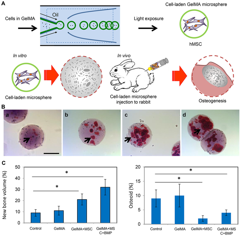
Figure 29.
Bone regeneration using cell-laden GelMA microspheres. (A) Schematic illustration for the fabrication of cell-laden GelMA microspheres using a photo-cross-linking-microfluidic method, and in vitro and in vivo applications for osteogenesis and bone regeneration in a rabbit model. (B) Alizarin red staining results of cell-laden GelMA microspheres after (a) 1, (b) 2, (c) 3, and (d) 4 weeks of culture for in vitro osteogenesis. Scale bar: 100 μm. (C) Histomorphometric results (%) of new bone (left) and osteoid (right) formation. Reprinted with permission from ref 1083. Copyright 2016 WILEY-VCH Verlag GmbH & Co. KGaA, Weinheim.

