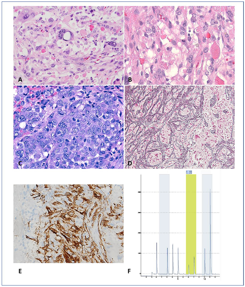
Figure 2. Anaplastic pleomorphic xanthastrocytoma (A-PXA) (WHO grade III): pathology and molecular genetic features.

A 40-year-old man presented with a temporal lobe mass. Histologic features included moderate cellularity, pleomorphism and numerous eosinophilic granular bodies (A). In addition, mitotic activity was easy to find (B). Cellular areas with a paradoxic decrease in pleomorphism and epithelioid morphology is a feature of some A-PXA (C). There is an increased in reticulin deposition (D) and CD34 expression (E) in this A-PXA. Detection of V600E by pyrosequencing assay (F). Analysis of sequence flanking the T>A hotspot (yellow shading) within codon 600 allows for the detection of V600E mutation. Nucleotide dispensation order and the numerical positions 5 and 10 are shown below the pyrogram.
