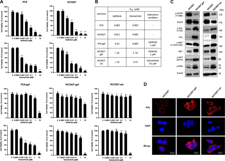
Fig. 1. Overexpression of AXL is observed in EGFR-TKI resistant NSCLC cells.
NSCLC cells (PC9, PC9-gef, HCC827, HCC827-gef, and HCC827-osi) were treated with gefitinib or osimertinib for 72 h. Cell viability was measured by MTT assay as described in Methods (a). IC50 values and subculture conditions were stated (b). The protein expressions of major RTKs, p-AKT, and p-ERK were determined by Western blotting (c). Cells were incubated on the confocal dish for 24 h, fixed, and stained with C-term AXL (red) and DAPI (blue) for visualization of cellular localization (d). Data are presented as the mean fold changes ± SD of three independent experiments. *P < 0.05, **P < 0.01, ***P < 0.005 by t-test

