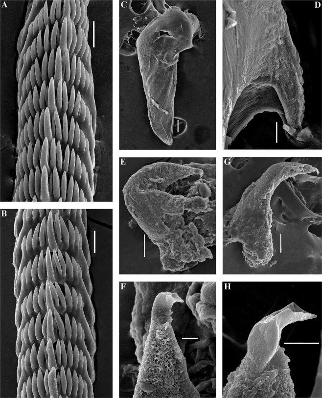
Figure 4.
Trinchesia caerulea (Montagu, 1804). Internal morphology, scanning electron microscopy. (A) Posterior part of radula of specimen from Spain, L’Estartit, Girona (ZMMU Op-648). (B) Posterior part of radula of specimen from Norway, Gulen Dive Resort (ZMMU Op-622). (C) Jaw of specimen from Norway (Gulen). (D) Details of masticatory processes of jaws, same specimen. (E) Copulative organ with stylet of specimen from Norway. (F) Stylet, details, same specimen. (G) Copulative organ with stylet of specimen from Spain. (H) Stylet details, same specimen. Scale bars: a, b, d −20 μm, c −100 μm, e, g −50 μm, f, h −10 μm. SEM micrographs: Alexander Martynov.

