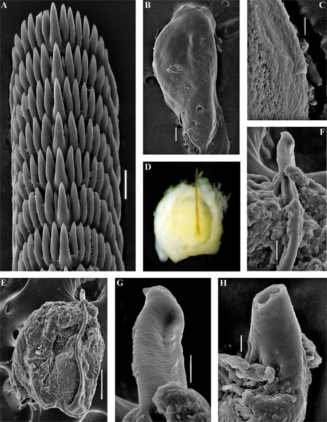
Figure 6.
Trinchesia cuanensis sp. n. Internal morphology, scanning and light electron microscopy. (A) Posterior part of radula of holotype from Northern Ireland (ZMMU Op-650). (B) Jaw, holotype. (C) Details of masticatory processes of jaws, holotype. (D) Copulative organ with stylet of paratype from Northern Ireland (GNM Gastropoda – 9054), light microscopy. (E) Same, scanning electron microscopy. (F) Details of stylet, same paratype. (G) Details of apical part of stylet, same specimen. (H) Cross section of basal part of stylet, showing channel inside, holotype. Scale bars: a, f −20 μm, b, e −100 μm, c, h, g −10 μm. SEM micrographs: Alexander Martynov.

