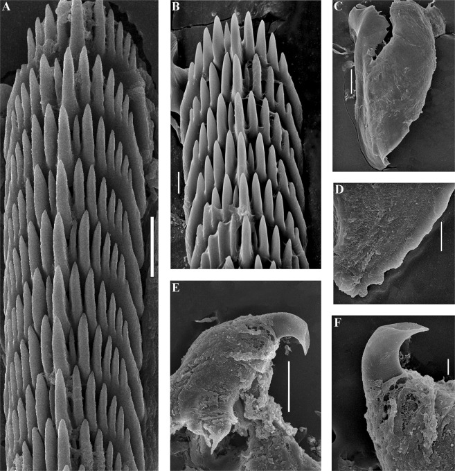
Figure 8.
Trinchesia morrowae sp. n. Internal morphology, scanning electron microscopy. (A) Posterior part of radula of paratype from Spain, L’Estartit (ZMMU Op-652). (B) Posterior part of radula of paratype from France, Banyuls-sur-Mer. (C) Jaw of specimen from Spain (L’Estartit, Girona). (D) Details of masticatory processes of jaws, same specimen. (E) Copulative organ with stylet of specimen from France, Banyuls-sur-Mer (ZMMU Op-653). (F) Details of stylet, paratype. Scale bars: (a) −20 μm; (b), (d), (f) −10 μm; (c) −100 μm; (e) −50 μm. SEM micrographs: Alexander Martynov.

