Figure 1.

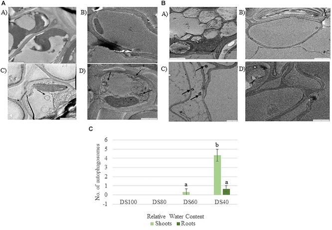
(A) Autophagosomes in Tripogon loliiformis shoots during dehydration. (A) Hydrated shoots (B) 80 RWC (C) 60 RWC (D) 40 RWC. (B) Few autophagosomes observed in T. loliiformis roots during dehydration. (A) Hydrated shoots (B) 80 RWC (C) 60 RWC (D) 40 RWC. (C) Comparison in autophagosome number between dehydrated T. loliiformis shoots and roots. P < 0.05. Samples denoted with the same letter were not statistically different from each other using a P-value < 0.05.
