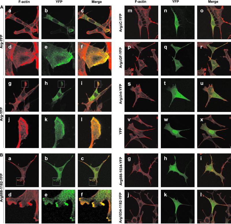
Figure 4.
Arg directs the formation of actin-rich structures at the lamellipodia of Swiss 3T3 cells. Swiss 3T3 cells were transfected with Arg, Arg deletion, or Arg fragment-YFP fusions, plated on coverslips, and fixed 24 h posttransfection. F-actin was visualized with rhodamine phalloidin. (A) Colocalization of Arg-YFP and Arg deletion-YFP fusions with F-actin. (a–l) Arg-YFP; (m–o) ArgΔC-YFP; (p and r) ArgΔGF-YFP; (s–u) ArgΔInt-YFP; (v–x) YFP alone. (d–f) Enlargements of the boxed areas indicated in a–c. (j–l) Enlargements of the boxed areas indicated in g–i. (B) Colocalization of Arg fragment-YFP fusions with F-actin. (a–f) Arg688–1182-YFP; (g–i) Arg688-1034-YFP; (j–l) Arg1034–1182-YFP. (d–f) Enlargements of the boxed areas indicated in a–c.

