Figure 3.
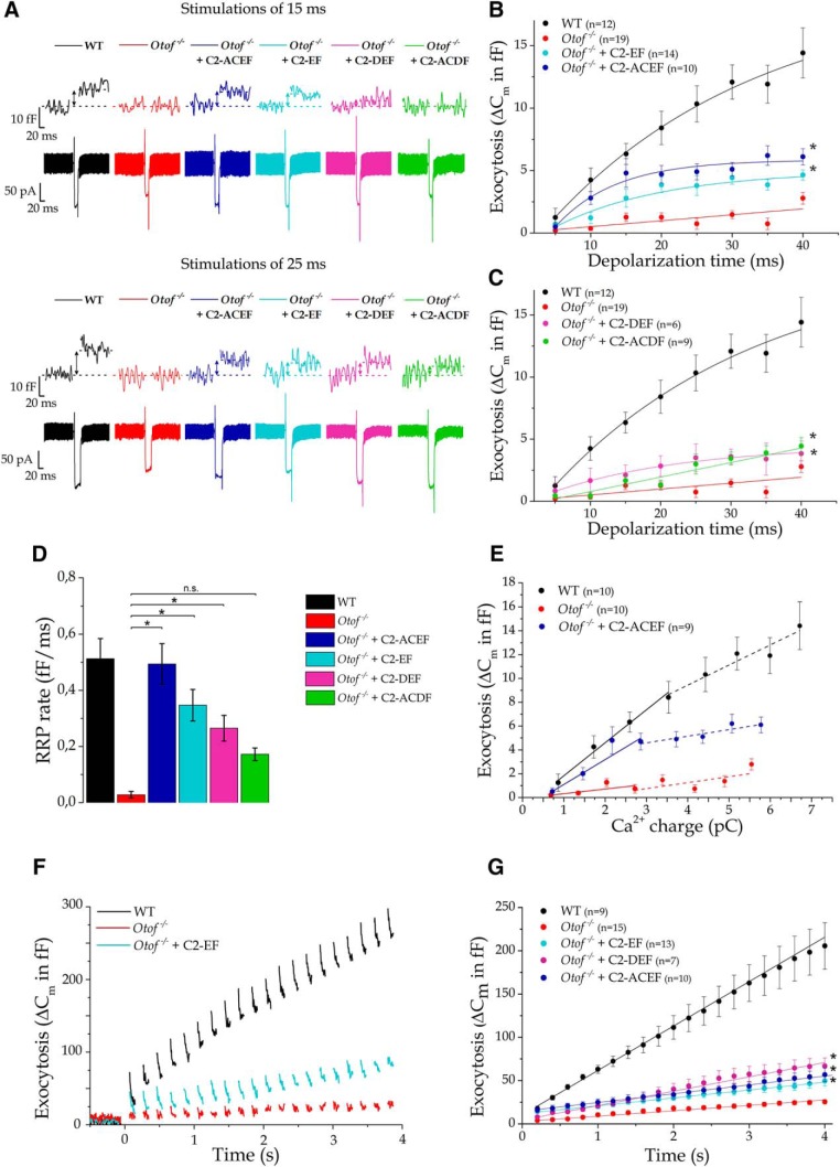
Mini-Otof constructs partially restore RRP exocytosis but not its vesicle resupply in P15–P21 Otof−/− mice. A, Example traces of vesicular exocytosis (ΔCm) and Ca2+ currents in IHCs triggered by 15 and 25 ms depolarizing steps from −80 to −10 mV in WT, Otof−/−, and C2-ACEF-, C2-EF-, C2-DEF-, and C2-ACDF-injected Otof−/− mice. B, Comparative kinetics of RRP exocytosis (depolarizing steps from −80 to −10 mV with increasing duration from 5 to 40 ms) for Otof-C2-ACEF-treated (dark blue) and C2-EF-treated (light-blue) Otof−/− IHCs. C, RRP kinetics in C2-DEF (purple) and C2-ACDF (green). For B and C, note a significant restoration of the exocytotic response compared with sham Otof−/− IHCs. *p < 0.05 (two-way ANOVA). Kinetics were best fitted with a single exponential with R2 > 0.94 except for Otof-C2-ACDF and Otof−/−. D, Comparative rate of RRP exocytosis obtained by fitting with a linear function the first four time–data points of the kinetics shown in Figure 4, B and C (linear slope in femtofarad per millisecond). The expression of the mini-Otof constructs C2-ACEF, C2-EF, and C2-DEF allowed a significant increase in the rate of exocytosis compared with sham-injected Otof−/−. *p < 0.05 (one-way ANOVA; see individual p values in Table 2). E, Ca2+ efficiency of RRP exocytosis (ΔCm/QCa) where the loading charge in Ca2+ (QCa) was obtained by integrating the Ca2+ current evoked during a voltage step from −80 to −10 mV for different brief pulses lasting 5, 10, 15, and 20 ms (full line). The slope of Otof-C2-ACEF IHCs was not significantly different of the WT slope (2.77 ± 0.21 and 2.09 ± 0.28 fF/pC, respectively; F test with F = 2.18 and p = 0.23), which is larger than in Otof−/− mice (0.39 ± 0.20 fF/pC; p = 5.8 10−3). For longer pulses (25, 30, 35, and 40 ms; dashed lines), exocytosis Ca2+ efficiency (ΔCm/QCa) in Otof-C2-ACEF IHCs is decreased, with a slope similar to that for noninjected Otof−/− mice (0.56 ± 0.14 and 0.50 ± 0.30 fF/pC, respectively; F test with F = 0.015 and p = 0.98). F, To evaluate the replenishment of the IHC active zone in vesicles, exocytosis was recorded during a train of 100 ms pulses from −80 to −10 mV, each stimulation separated by 100 ms. Examples of exocytotic traces of vesicular recruitment in control WT, Otof−/−, and Otof-C2-EF-treated IHCs. G, Comparative mean recruitment in Otof-C2-EF-, Otof-C2-DEF-, and Otof-C2-ACEF-treated IHCs. Note that in Otof−/− IHCs (red), the vesicles recruitment is severely impaired. When mini-Otof constructs are expressed in Otof−/− IHCs, the vesicle supply is only slightly, but significantly, restored compared to Otof−/− (two-way ANOVA, p = 4.3 10−8; Tukey's post hoc test, p < 10−4 for each as indicated by the asterisks), except for Otof-C2-ACDF (data not shown; p = 0.16).

