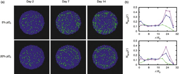
Figure 5.

Simulated chondrogenic differentiation of mesenchymal stem cells in pellet culture at 20% pO2. (a) Spheroids of cells expanded at 5% pO2 (upper row) and at 20% pO2 (lower row). Functional differentiated chondrocytes are shown in green, other cells in blue. (b) Fraction of chondrocytes versus radial distance from the spheroid centre at days 2 (green), 7 (blue) and 14 (magenta). At day 14, the total number of chondrocytes in pellets of cells expanded at 5% pO2 is about 1.5 times larger then in pellets of cells expanded at 20% pO2.
