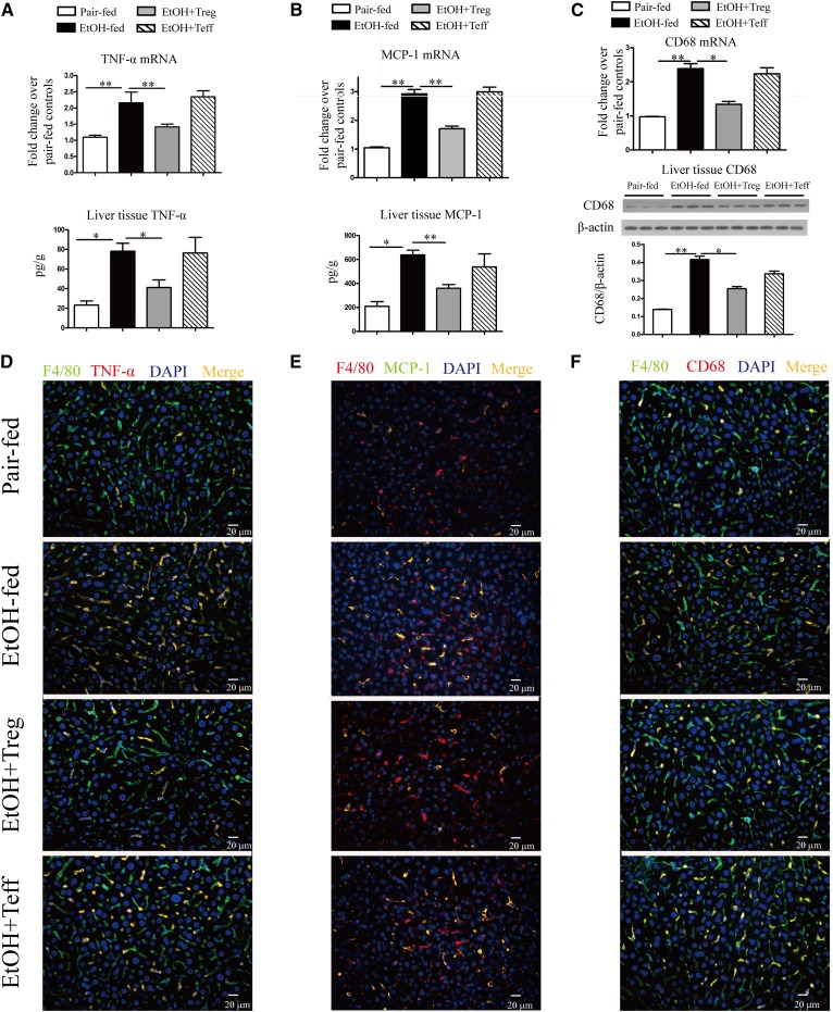
Fig. 6.
Adoptive transfer of Tregs prevents hepatic MCP-1 and TNF-α overexpression in AFL in mice. C57BL/6 mice were fed a 5% alcohol-containing Leiber-DeCarli diet or isocaloric pair-fed diet for 6 weeks. A: Livers were subjected to analysis of TNF-α mRNA expression was by real-time PCR, and liver tissue TNF-α levels were estimated by ELISA (*P < 0.05, **P < 0.01; n = 8). B: MCP-1 mRNA by real-time PCR and liver tissue MCP-1 by ELISA (*P < 0.05, **P < 0.01; n = 8). C: CD68 mRNA was estimated by real-time PCR (*P < 0.05, **P < 0.01; n = 8). Liver tissue CD68 levels by Western blotting are shown in representative gels (upper panel) and graphs of density units (lower panel). D: Representative sections of TNF-α immunofluorescence staining (magnification, ×400; green, F4/80; red, TNF-α; yellow, F4/80 and TNF-α double positive). E: Representative sections of MCP-1 immunofluorescence staining (magnification, ×400; red, F4/80; green, MCP-1; yellow, F4/80 and MCP-1 double positive). F: Representative sections of CD68 immunofluorescence staining (magnification, ×400; green, F4/80; red, CD68; yellow, F4/80 and CD68 double positive). EtOH, ethanol.

