
Figure 1.
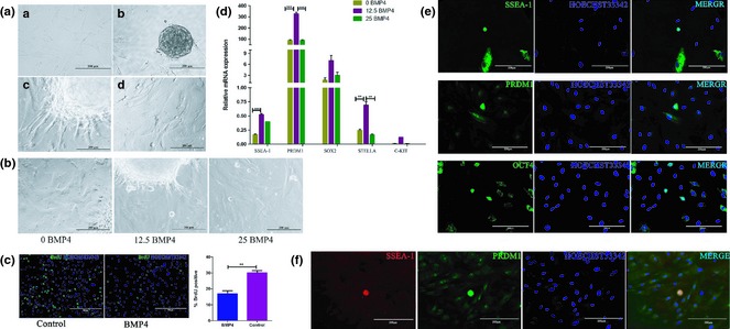
BMP4 effect on differentiation of hUC ‐MSCs into PGC‐like cells. (a) Morphology of hUC‐MSCs under induction of BMP4. a. Untreated hUC‐MSCs cultured in normal culture medium. b. EBs were formed from hUC‐MSCs. c. Adherent EB before induction. d. Cell morphology after 7 days induction. (b) After 7 days induction with BMP4, round‐shaped cells migrated out from EBs. (c) Proliferation profile of BMP4 induced cells or untreated cells was determined by BrdU incorporation assay. (d) qRT‐PCR analysis examined relative expression levels of SSEA‐1, C‐KIT, PRDM1, SOX2 and STELLA. These PGC‐specific markers were expressed at higher levels in the 12.5 ng/ml BMP4‐induced group. (e) Immunofluorescence staining for SSEA‐1, PRDM1 and OCT4. Induced round‐shaped cells highly expressed these PGC‐specific markers and had higher nucleus‐cytoplasmic ratio. (f) Co‐staining for SSEA‐1 and PRDM1. Round‐shaped cells were double‐positive for these two markers.