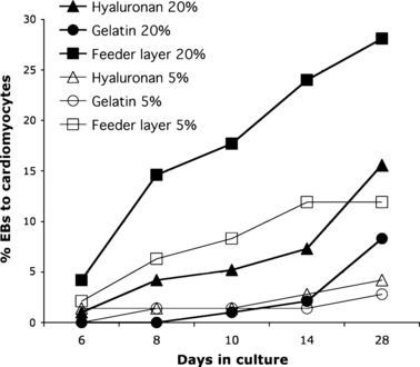
Figure 8.

Beating cardiomyocyte formation over 30 days. EBs from mES cells were cultured under different conditions of oxygen concentrations (20%, filled symbols, or 5%, open symbols) and substrata – 120 μg/μl hyaluronan (triangles), 0.1% gelatin (circles) and mEF (squares) – and rates of beating cardiomyocytes counted at indicated times. Under hypoxic conditions, levels of differentiation into beating cardiomyocytes were significantly lower than under normoxic conditions on feeder layer (P = 0.014) and on HA (P = 0.035). Under normoxic conditions, feeder layers supported significantly higher cardiomyocyte formation when compared to 0.1% gelatin (P < 0.001) or to HA (P = 0.05).
