
Fig. 3.
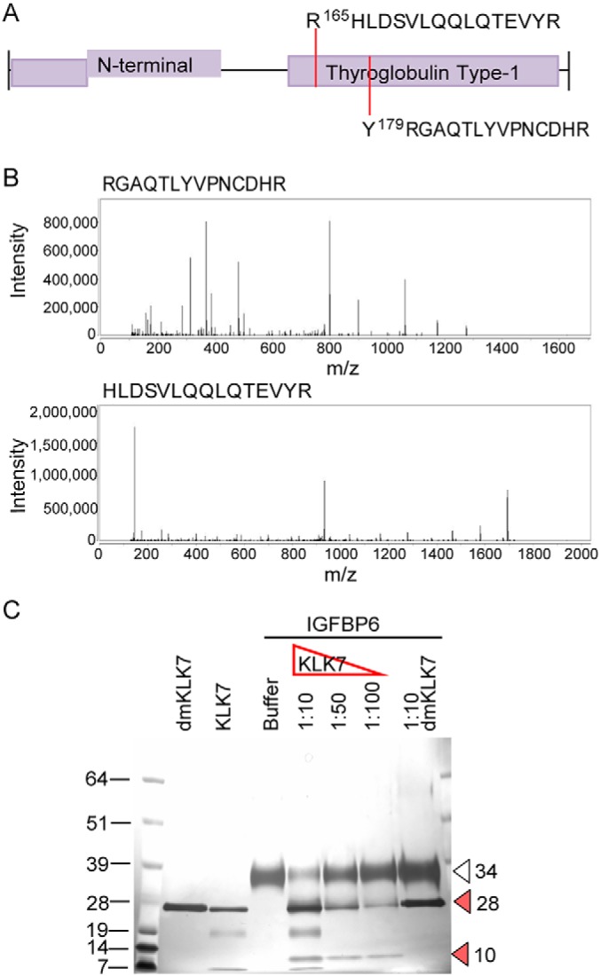
KLK7-mediated hydrolysis of IGFBP6 in SKOV-3 and OVMZ-6 cell CM. A, TAILS-identified cleavage sites are indicated by dotted lines on the schematic of selected protein domains, based on annotation in the UniProtKB. B, Respective spectra of the TAILS-identified peptides. C, Silver-stained 12% SDS-PAGE showing hydrolysis of recombinant IGFBP6. Arrowheads indicate: open: full length IGFBP6, 34 kDa; red: cleaved IGFBP6 fragments, 10 and 28 kDa. The molecular weight (MW) of the protein standard (kDa) is indicated to the left.