Fig. 5.
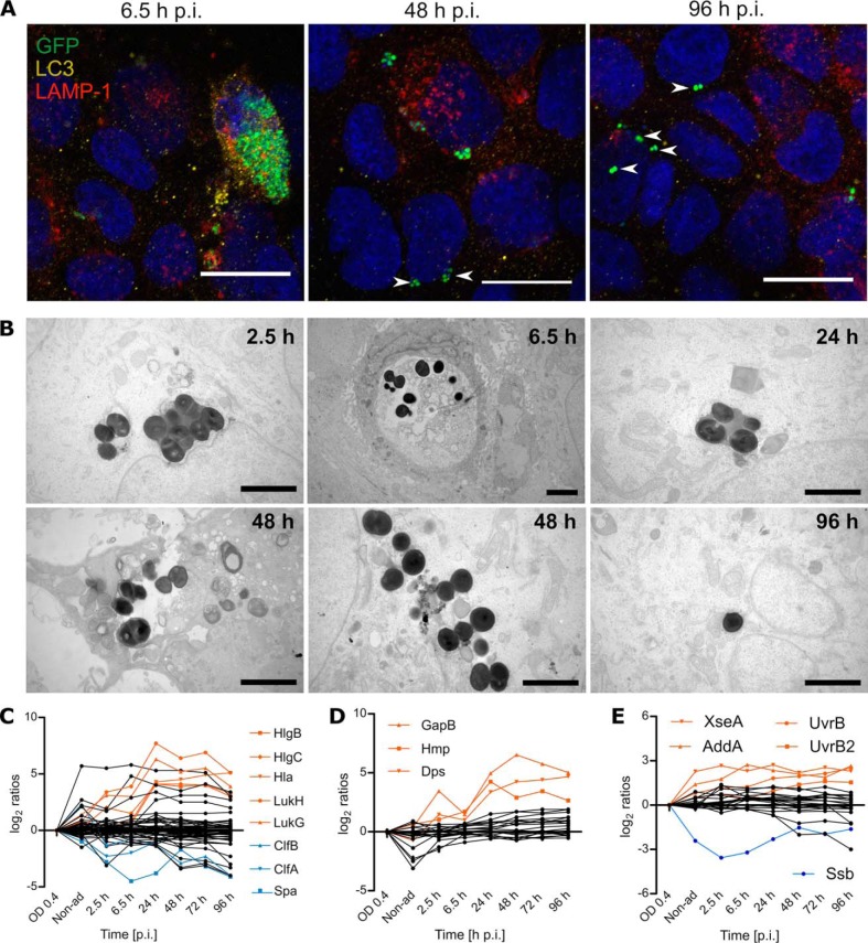
The persistent subpopulation of S. aureus is mostly found in the cytosolic environment of the host. Intracellular localization of S. aureus was examined by colocalization with the protein markers LC3 for phagosomes, and LAMP-1 for lysosomes (A; scale bar: 20 μm). After internalization of S. aureus by the HBE cells, most of the bacterial population is located inside closed compartments. Still, the percentage of bacteria that escapes the vesicles (white arrow heads) increases over time and most of the bacteria are found in a cytosolic environment by the end of the infection. These results were corroborated by electron microscopy (B; scale bar = 2 μm). During the first hours of infection most bacteria are located inside degradative vesicles (dark compartments) or phagosomes (light compartments), but single bacteria escaped the closed compartments as early as 2.5 h p.i. (supplemental Fig. S4). By the end of the time of observation, at 72 h and 96 h p.i., all observed bacterial clusters are cytosolic. The displayed images are representative of different time points, additional images are provided in supplemental Figs. S3 and S4. The escape from the compartments could be induced by proteins related to pathogenesis, including toxins regulated by Agr and SaeRS (C). Moreover, this escape might have an impact on the production of stress proteins related to oxidative stress (D) and DNA repair (E). The list of proteins included in the line plots is available in the supplemental Table S8. A selection of significantly (p value<0.01) regulated proteins is displayed in orange and blue. The mass spectrometry data represents the average of four biological replicates.

