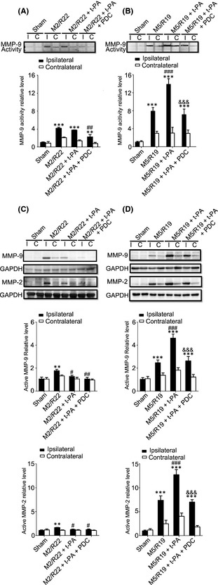
Figure 4.

Effect of PDC (FeTMPyP, 3 mg/kg) on MMP‐9 activity and MMP‐9/MMP‐2 expression in MCAO ischemic rat brains with early and delayed t‐PA treatments. Rats were treated with t‐PA (10 mg/kg) at 2 and 5 h of MCAO ischemia following 22 and 19 h of reperfusion, respectively. M2/R22, 2 h of MCAO plus 22 h of reperfusion; M5/R19, 5 h of MCAO plus 19 h of reperfusion. I, ipsilateral; C, contralateral. (A) Representative gelatin zymography and statistical results in M2/M22 groups (ipsilateral) (**P < 0.01, ***P < 0.001, compared to sham control; ## P < 0.01, compared to M2/R22). (B) Representative gelatin zymography and statistical results in M5/M19 groups (ipsilateral) (***P < 0.001, compared to sham control; ### P < 0.001, compared to M5/R19; &&& P < 0.001, compared to M5/R19 + t‐PA). (C) Representative Western blot and statistical analysis for MMP‐9/MMP‐2 expressions in M2/R22 groups (**P < 0.01, compared to sham control; # P < 0.05, ## P < 0.01, compared to M2/R22). (D) Representative Western blot and statistical analysis for MMP‐9/MMP‐2 expressions in M5/R19 groups (***P < 0.001, compared to sham control; ### P < 0.001, compared to M5/R19; &&& P < 0.001, compared to M5/R19 + t‐PA). Data are expressed as mean ± SEM (n = 3–4).
