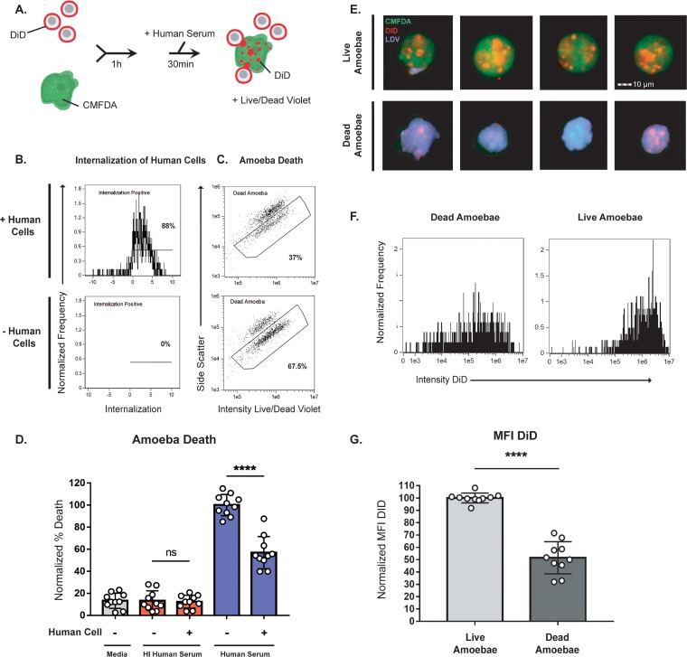
FIG 3.
Interaction with human cells leads to protection from lysis by human serum. (A) CMFDA-labeled amoebae were incubated alone or in the presence of DiD-labeled human cells for 1 h. Cells were then exposed to either active human serum, heat-inactivated human serum, or M199s medium. Following exposure to serum, samples were stained with Live/Dead violet, and viability was quantified using imaging flow cytometry, with 10,000 images collected for each sample. (B) Representative plots showing internalization of human cells from amoebae incubated with human cells or in the absence of human cells. (C) Representative plots comparing amoebic death from the conditions shown in panel B. (D) Quantification of amoebic death for all experimental conditions. Cells exposed to M199s medium are shown in gray, to heat-inactivated (HI) human serum in red, and to active human serum in blue. Percentages of dead amoebae were normalized to numbers of dead amoebae in the amoeba-alone samples that were treated with active human serum. (E) Representative images of live and dead amoebae from amoebae coincubated with human cells and exposed to active human serum. Amoebae are shown in green, human cell membranes in red, and dead cells in violet. (F) Representative histograms showing the mean fluorescence intensity (MFI) of DiD in live and dead amoebae from samples exposed to human serum. (G) Quantification of the DID MFI shown in panel F. Ten replicates across 5 independent experiments were performed. ns, not significant.

