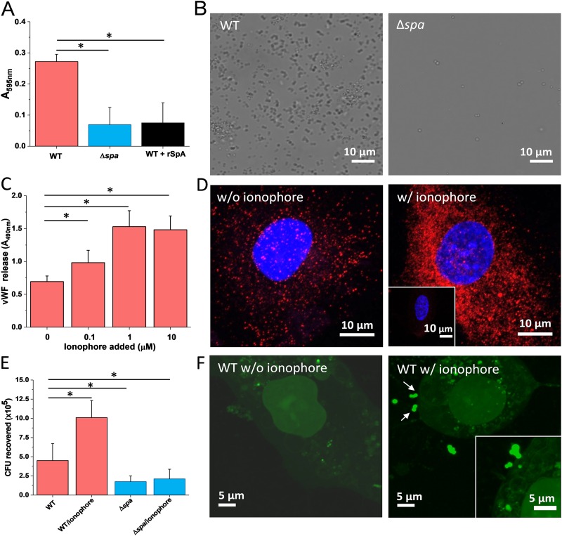
FIG 1.
Attachment of S. aureus to immobilized vWF and to endothelial cells. (A) Microtiter wells coated with vWF were incubated with S. aureus Newman WT and Newman Δspa bacteria, rinsed, and stained with crystal violet, and the absorbance at 595 nm was measured in an ELISA plate reader. Attachment of WT bacteria was also performed in the presence of 2 μg soluble SpA. (B) Optical microscopy images of S. aureus Newman WT and Newman Δspa bacteria adhering to vWF-coated substrates. (C) vWF release by endothelial cells. Confluent HUVEC monolayers were incubated with increasing concentrations of calcium ionophore A23187 for 10 min. The vWF released in the extracellular matrix was determined with rabbit anti-vWF polyclonal antibody followed by HRP-conjugated goat anti-rabbit. (D) Imaging of vWF release. Shown are confocal microscopy images of HUVECs before (left) and after (right) treatment with calcium ionophore. Staining with mouse anti-vWF antibody and secondary goat anti-mouse Alexa Fluor 647 antibody documents the release of large amounts of vWF upon ionophore treatment (vWF is in red, and the nucleus is in blue). The inset shows a control experiment in which the primary antibody was missing. (E) Endothelial cell adhesion assays. Confluent HUVECs were incubated with S. aureus Newman WT and Newman Δspa bacteria for 90 min, in the presence or absence of 1 μM ionophore. The number of adhering bacteria was determined as described in Materials and Methods. (F) Imaging of bacterial-endothelial cell adhesion. Confluent HUVECs, treated (right) or not (left) with ionophore, were incubated with S. aureus Newman WT bacteria stained with the BacLight viability kit, rinsed, and imaged by confocal microscopy. The arrows indicate bacteria adhering to the endothelial cell surface. For all plots, means and SD of results from two independent experiments, each performed in triplicate, are presented. Statistically significant difference is indicated (Student's t test; *, P < 0.05).

