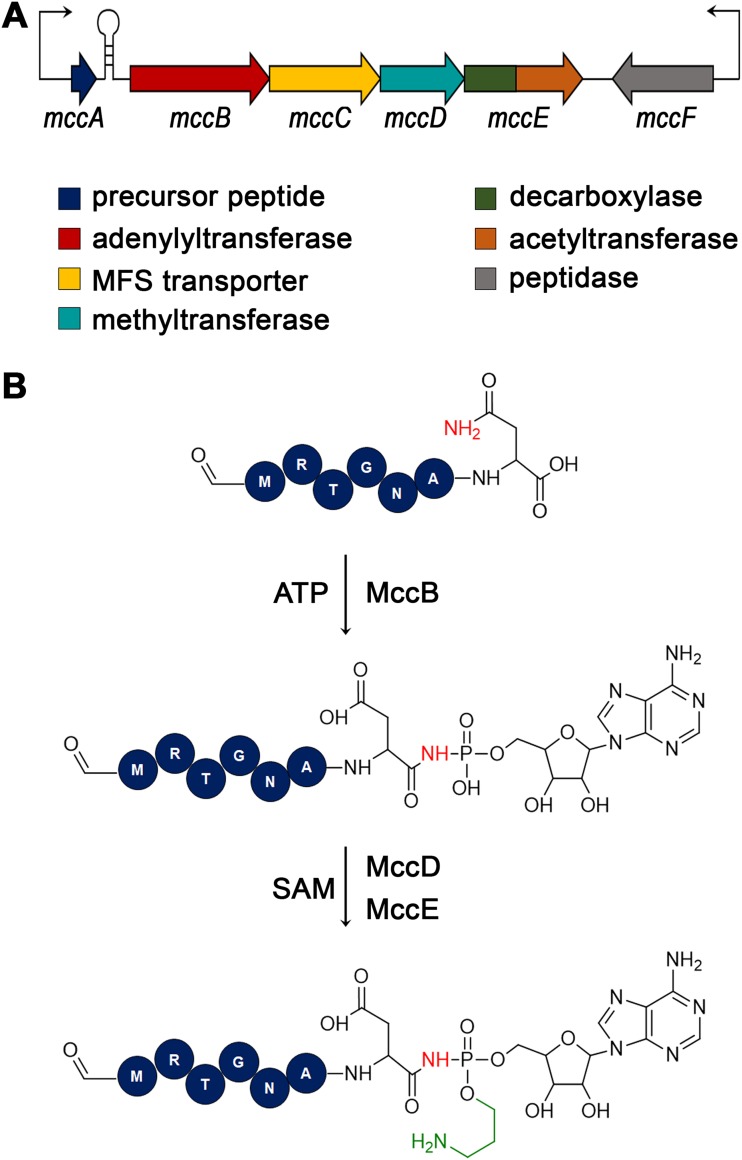
FIG 1.
Escherichia coli mcc gene cluster and biosynthesis of microcin C. (A) The E. coli mcc biosynthetic gene cluster is schematically shown. Genes are shown by colored arrows and the functions of gene products are indicated below. Thin arrows indicate promoters from which transcription of mcc genes is initiated. A transcription terminator located between the mccA and mccB genes is shown as a hairpin. (B) The steps of the McC biosynthesis pathway and enzymes involved are presented. For the peptide part, the first 6 amino acids are shown as circles with their identity indicated in a single-letter amino acid code. The last amino acid is shown as a skeletal formula. The N-terminal methionine residue of mature McC is formylated.

