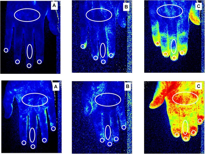
FIGURE 1.

Laser Speckle Contrast Analysis (LASCA) images of secondary Raynaud’s phenomenon (RP) to systemic sclerosis, in a patient with a “Late” pattern of scleroderma microangiopathy (A), primary RP (B) and a healthy subject (C), showing the regions of interest (ROI - white circles) created at the level of dorsum and palm of the hand, dorsal and palmar aspect of the 3rd finger, periungual areas and fingertips to evaluate blood perfusion. Color code: blue corresponds to a low BP, yellow an intermediate BP and red a higher BP. Noteworthy is the fact that subjects with a late pattern have a prevalence of blue, indicating a low perfusion level.
