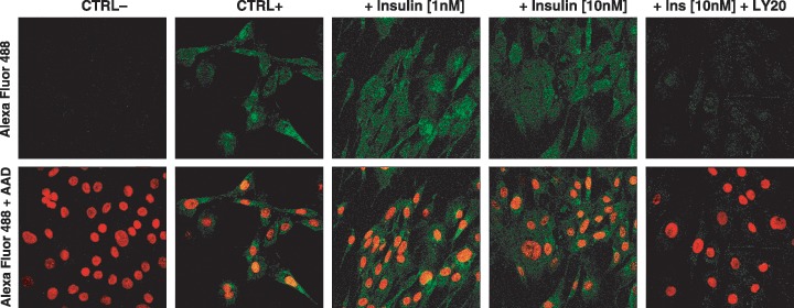
Figure 6.

Images from confocal microscopy showing cytoplasmic and nuclear localization of myogenin protein in cell cultures of C2C12 muscle cells on the third day of experiment. Top panel: insulin induces myogenin protein expression in C2C12 muscle cells. Bottom panel: merged pictures of the same visual fields with nuclear DNA stained with AAD within the cells. From left to right: negative control (without primary antiserum, CTRL–), positive control (without insulin, CTRL+), effect of insulin (1 nm), effect of insulin (10 nm), effect of LY294002 (20 µm) being present 30 min prior to insulin (10 nm) addition. Images were gathered as single slides under 40× lens and 5× digital zoom. Typical results from two experiments are shown.
